Flash sale: From now to October 31st, 2019
Promotion product list: Classical loading control and tag antibodies and the conjugated versions of these antibodies, including HRP, Biotin, FITC, Cy3, Cy5 and AbFlour TM dyes.
Details of activities: Buy 2 get 1 for free (if a single order is full of 3 antibodies, the antibody with the lowest price will be exempted).
Why choose Abbkine loading control and tag antibodies?
- Abbkine offers a wide range of
loading control and tag antibodies, including the hot targets and unique
species, covering the molecular weights from 15 kDa to 116 kDa. - Abbkine conjugated loading
control and tag antibodies have diverse conjugates, including enzyme and
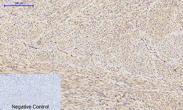
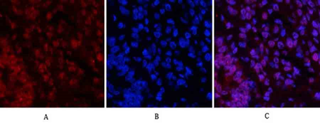
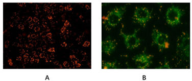
featured fluorescent dyes, with the application of WB, IHC, IF and IP
experiments. - Abbkine loading control and
tag antibodies are verified through rigorous procedures, popular with customers
for their good performance and high sensitivity. - Different sizes for clients to
choose, small size and bulk size are available, both with high
cost-effectiveness.
Part of promotion product list:
Classical loading control and tag antibodies (Citation of over 100 SCI Documents)
Cat# | Product name | Application | size |
A01010 | Anti-β-Actin Mouse Monoclonal Antibody (1C7) | WB#IHC-P#IF | 50/200/200x5ul |
A01020 | Anti-GAPDH Mouse Monoclonal Antibody (2B5) | WB#IHC-P#IF | 50/200/200x5ul |
A01030 | Anti-β-Tubulin Mouse Monoclonal Antibody (3G6) | WB#IHC-P#IF | 50/200/200x5ul |
A01050 | Anti-Plant Actin Mouse Monoclonal Antibody (3T3) | WB | 50/200/200x5ul |
A02010 | Anti-DDDDK Tag Mouse Monoclonal Antibody (1B10) | WB#IF#IP | 50/200/200x5ul |
A02050 | Anti-His Tag Mouse Monoclonal Antibody (5C3) | WB#IF#IP | 50/200/200x5ul |
A02040 | Anti-HA Tag Mouse Monoclonal Antibody (4F6) | WB#IF#IP | 50/200/200x5ul |
A02020 | Anti-GFP Tag Mouse Monoclonal Antibody (3D3) | WB#IP | 50/200/200x5ul |
A02030 | Anti-GST Tag Mouse Monoclonal Antibody (2A8) | WB | 50/200/200x5ul |
A02060 | Anti-Myc Tag Mouse Monoclonal Antibody (2D5) | WB#IF#IP | 50/200/200x5ul |
A02080 | Anti-mCherry Tag Mouse Monoclonal Antibody (9D3) | WB#IF | 50/200/200x5ul |
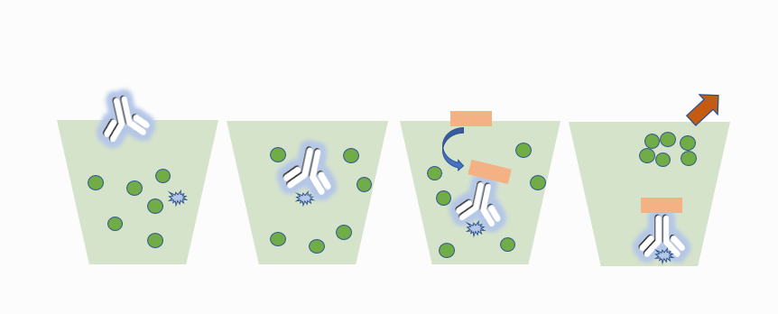
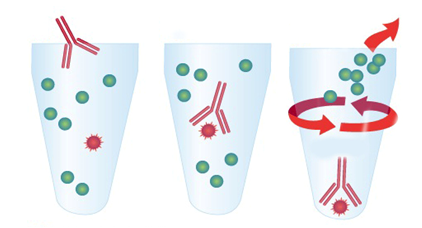
IP experiment partner-Agarose/Magnetic beads conjugated tag antibodies
Compared
with the traditional immunoprecipitation (IP) method using Protein A/G,
Abbkine's agarose/magnetic bead coupling labeled antibody eliminates the step
of Protein A/G binding with antigen-antibody complex. After the conjugated
antibody is directly combined with the target protein, the target protein is
separated from the cell lysate by centrifugation or using a magnetic rack. It
not only makes the experiment simple and fast, but also can avoid non-specific
binding and reduce the background.
Cat# | Product name | Application | size |
A02010AGB | Anti-DDDDK Tag Mouse Mab (1B10), Agarose | IP | 100ul/400ul/2ml |
A02010MGB | Anti-DDDDK Tag Mouse Mab (1B10), Magnetic Beads | IP | 100ul/400ul/2ml |
A02040AGB | Anti-HA Tag Mouse Mab (4F6), Agarose | IP | 100ul/400ul/2ml |
A02040MGB | Anti-HA Tag Mouse Mab (4F6), Magnetic Beads | IP | 100ul/400ul/2ml |
A02050AGB | Anti-His Tag Mouse Mab (5C3), Agarose | IP | 100ul/400ul/2ml |
A02050MGB | Anti-His Tag Mouse Mab (5C3), Magnetic Beads | IP | 100ul/400ul/2ml |
A02060AGB | Anti-Myc Tag Mouse Mab (2D5), Agarose | IP | 100ul/400ul/2ml |
A02060MGB | Anti-Myc Tag Mouse Mab (2D5), Magnetic Beads | IP | 100ul/400ul/2ml |
A02170AGB | Anti-V5 Tag Mouse Mab (11D5), Agarose | IP | 100ul/400ul/2ml |
A02170MGB | Anti-V5 Tag Mouse Mab (11D5), Magnetic Beads | IP | 100ul/400ul/2ml |
A02180AGB | Anti-VSV-G-Tag Mouse Mab (14D2), Agarose | IP | 100ul/400ul/2ml |
A02180MGB | Anti-VSV-G-Tag Mouse Mab (14D2), Magnetic Beads | IP | 100ul/400ul/2ml |
For
the full product promotion list, please contact Abbkine's local distributor or
contact service@abbkine.com directly. Abbkine has the final right to interpret
this activity.