2019年8月21日星期三

Selection Strategy of Apoptosis Products- Early apoptosis

Apoptosis
is a dynamic process, which involves a series of complex biochemical reactions,
expression regulation of multiple genes, signal transduction, cascade reactions
involving multiple enzymes and multiple signal pathways. The cell feels the
corresponding apoptosis signal stimulation, and a series of control switches in
the cell are turned on or off, and activation of various enzymes triggers a
series of cascade reactions. Different external factors initiate apoptosis in
different ways, resulting in different signal transduction.


Early apoptosis


Feature 1: eversion of lipid membrane
inside


In normal cells, phosphatidylserine (PS) is only distributed on the inner
side of the lipid bilayer of the cell membrane. In the early stage of cell
apoptosis, PS turns from the inner side of the lipid membrane to the outer
side. Annexin V is a calcium-dependent phospholipid binding protein with a
molecular weight of 35-36kD, and has high affinity with PS. It binds to the
cell membrane of early apoptotic cells through PS exposed on the outside of
cells. Therefore Annexin V is a sensitive indicator to detect early apoptosis
of cells. Usually, Annexin V is labeled with some fluorescent probes, which can
simply and directly detect PS eversion, an important feature of apoptosis.


Product recommendation


Product name

Cat#

Abbkine Annexin
V-AbFluor™ 405 Apoptosis Detection Kit

KTA0001

Abbkine Annexin
V-AbFluor™ 488 Apoptosis Detection Kit

KTA0002

Abbkine Annexin
V-AbFluor™ 555 Apoptosis Detection Kit

KTA0003

Abbkine Annexin
V-AbFluor™ 647 Apoptosis Detection Kit

KTA0004


Product character


AbFlourTM
series of characteristic fluorescent dyes (blue, green and red) patented by
Abbkine label recombinant human Annexin V protein, and are matched with PI to
introduce different fluorescence apoptosis detection kits, which are suitable
for detection with fluorescence equipment such as flow cytometry and
fluorescence microscope.


Feature 2: Mitochondrial membrane
potential decreased


Normal mitochondrial membrane potential is the premise for maintaining
mitochondrial oxidative phosphorylation and ATP production, and is necessary
for maintaining mitochondrial function. The decline of mitochondrial membrane
potential is a landmark event in the early stage of cell apoptosis. JC-1 is an
ideal fluorescent probe widely used for detecting mitochondrial membrane
potential. When mitochondrial membrane potential is high, JC-1 aggregates in
the matrix of mitochondria to form polymers (J-aggregates). Red fluorescence
can be generated(Ex/Em =
585/590). when the mitochondrial membrane potential is low, JC-1 cannot
aggregate in the matrix of mitochondria. at this time, JC-1 is a monomer, which
can generate green fluorescence (Ex/Em = 510/527 nm).


Product recommendation


Product name

Cat#

Mitochondrial Membrane Potential Assay Kit
(JC-1)

KTA4001


Product character


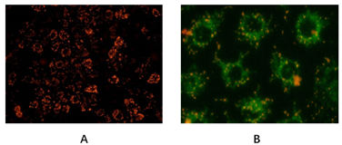
Abbkine Mitochondrial Membrane Potential Analysis Kit (JC-1) uses JC-1 as a fluorescent probe to rapidly and sensitively detect changes in mitochondrial membrane potential. This kit can very conveniently detect changes in mitochondrial membrane potential through changes in fluorescence color.


Abbkine Mitochondrial Membrane Potential Analysis Kit (JC-1)

Selection Strategy of Apoptosis Products- Early apoptosis

Apoptosis
is a dynamic process, which involves a series of complex biochemical reactions,
expression regulation of multiple genes, signal transduction, cascade reactions
involving multiple enzymes and multiple signal pathways. The cell feels the
corresponding apoptosis signal stimulation, and a series of control switches in
the cell are turned on or off, and activation of various enzymes triggers a
series of cascade reactions. Different external factors initiate apoptosis in
different ways, resulting in different signal transduction.


Early apoptosis


Feature 1: eversion of lipid membrane
inside


In normal cells, phosphatidylserine (PS) is only distributed on the inner
side of the lipid bilayer of the cell membrane. In the early stage of cell
apoptosis, PS turns from the inner side of the lipid membrane to the outer
side. Annexin V is a calcium-dependent phospholipid binding protein with a
molecular weight of 35-36kD, and has high affinity with PS. It binds to the
cell membrane of early apoptotic cells through PS exposed on the outside of
cells. Therefore Annexin V is a sensitive indicator to detect early apoptosis
of cells. Usually, Annexin V is labeled with some fluorescent probes, which can
simply and directly detect PS eversion, an important feature of apoptosis.


Product recommendation


Product name

Cat#

Abbkine Annexin
V-AbFluor™ 405 Apoptosis Detection Kit

KTA0001

Abbkine Annexin
V-AbFluor™ 488 Apoptosis Detection Kit

KTA0002

Abbkine Annexin
V-AbFluor™ 555 Apoptosis Detection Kit

KTA0003

Abbkine Annexin
V-AbFluor™ 647 Apoptosis Detection Kit

KTA0004


Product character


AbFlourTM
series of characteristic fluorescent dyes (blue, green and red) patented by
Abbkine label recombinant human Annexin V protein, and are matched with PI to
introduce different fluorescence apoptosis detection kits, which are suitable
for detection with fluorescence equipment such as flow cytometry and
fluorescence microscope.


Feature 2: Mitochondrial membrane
potential decreased


Normal mitochondrial membrane potential is the premise for maintaining
mitochondrial oxidative phosphorylation and ATP production, and is necessary
for maintaining mitochondrial function. The decline of mitochondrial membrane
potential is a landmark event in the early stage of cell apoptosis. JC-1 is an
ideal fluorescent probe widely used for detecting mitochondrial membrane
potential. When mitochondrial membrane potential is high, JC-1 aggregates in
the matrix of mitochondria to form polymers (J-aggregates). Red fluorescence
can be generated(Ex/Em =
585/590). when the mitochondrial membrane potential is low, JC-1 cannot
aggregate in the matrix of mitochondria. at this time, JC-1 is a monomer, which
can generate green fluorescence (Ex/Em = 510/527 nm).


Product recommendation


Product name

Cat#

Mitochondrial Membrane Potential Assay Kit
(JC-1)

KTA4001


Product character


Abbkine Mitochondrial Membrane Potential Analysis Kit (JC-1) uses JC-1 as a fluorescent probe to rapidly and sensitively detect changes in mitochondrial membrane potential. This kit can very conveniently detect changes in mitochondrial membrane potential through changes in fluorescence color.


Abbkine Mitochondrial Membrane Potential Analysis Kit (JC-1)

Selection Strategy of Apoptosis Products- Early apoptosis

Apoptosis
is a dynamic process, which involves a series of complex biochemical reactions,
expression regulation of multiple genes, signal transduction, cascade reactions
involving multiple enzymes and multiple signal pathways. The cell feels the
corresponding apoptosis signal stimulation, and a series of control switches in
the cell are turned on or off, and activation of various enzymes triggers a
series of cascade reactions. Different external factors initiate apoptosis in
different ways, resulting in different signal transduction.


Early apoptosis


Feature 1: eversion of lipid membrane
inside


In normal cells, phosphatidylserine (PS) is only distributed on the inner
side of the lipid bilayer of the cell membrane. In the early stage of cell
apoptosis, PS turns from the inner side of the lipid membrane to the outer
side. Annexin V is a calcium-dependent phospholipid binding protein with a
molecular weight of 35-36kD, and has high affinity with PS. It binds to the
cell membrane of early apoptotic cells through PS exposed on the outside of
cells. Therefore Annexin V is a sensitive indicator to detect early apoptosis
of cells. Usually, Annexin V is labeled with some fluorescent probes, which can
simply and directly detect PS eversion, an important feature of apoptosis.


Product recommendation


Product name

Cat#

Abbkine Annexin
V-AbFluor™ 405 Apoptosis Detection Kit

KTA0001

Abbkine Annexin
V-AbFluor™ 488 Apoptosis Detection Kit

KTA0002

Abbkine Annexin
V-AbFluor™ 555 Apoptosis Detection Kit

KTA0003

Abbkine Annexin
V-AbFluor™ 647 Apoptosis Detection Kit

KTA0004


Product character


AbFlourTM
series of characteristic fluorescent dyes (blue, green and red) patented by
Abbkine label recombinant human Annexin V protein, and are matched with PI to
introduce different fluorescence apoptosis detection kits, which are suitable
for detection with fluorescence equipment such as flow cytometry and
fluorescence microscope.


Feature 2: Mitochondrial membrane
potential decreased


Normal mitochondrial membrane potential is the premise for maintaining
mitochondrial oxidative phosphorylation and ATP production, and is necessary
for maintaining mitochondrial function. The decline of mitochondrial membrane
potential is a landmark event in the early stage of cell apoptosis. JC-1 is an
ideal fluorescent probe widely used for detecting mitochondrial membrane
potential. When mitochondrial membrane potential is high, JC-1 aggregates in
the matrix of mitochondria to form polymers (J-aggregates). Red fluorescence
can be generated(Ex/Em =
585/590). when the mitochondrial membrane potential is low, JC-1 cannot
aggregate in the matrix of mitochondria. at this time, JC-1 is a monomer, which
can generate green fluorescence (Ex/Em = 510/527 nm).


Product recommendation


Product name

Cat#

Mitochondrial Membrane Potential Assay Kit
(JC-1)

KTA4001


Product character


Abbkine Mitochondrial Membrane Potential Analysis Kit (JC-1) uses JC-1 as a fluorescent probe to rapidly and sensitively detect changes in mitochondrial membrane potential. This kit can very conveniently detect changes in mitochondrial membrane potential through changes in fluorescence color.


Abbkine Mitochondrial Membrane Potential Analysis Kit (JC-1)

Selection Strategy of Apoptosis Products- Early apoptosis

Apoptosis
is a dynamic process, which involves a series of complex biochemical reactions,
expression regulation of multiple genes, signal transduction, cascade reactions
involving multiple enzymes and multiple signal pathways. The cell feels the
corresponding apoptosis signal stimulation, and a series of control switches in
the cell are turned on or off, and activation of various enzymes triggers a
series of cascade reactions. Different external factors initiate apoptosis in
different ways, resulting in different signal transduction.


Early apoptosis


Feature 1: eversion of lipid membrane
inside


In normal cells, phosphatidylserine (PS) is only distributed on the inner
side of the lipid bilayer of the cell membrane. In the early stage of cell
apoptosis, PS turns from the inner side of the lipid membrane to the outer
side. Annexin V is a calcium-dependent phospholipid binding protein with a
molecular weight of 35-36kD, and has high affinity with PS. It binds to the
cell membrane of early apoptotic cells through PS exposed on the outside of
cells. Therefore Annexin V is a sensitive indicator to detect early apoptosis
of cells. Usually, Annexin V is labeled with some fluorescent probes, which can
simply and directly detect PS eversion, an important feature of apoptosis.


Product recommendation


Product name

Cat#

Abbkine Annexin
V-AbFluor™ 405 Apoptosis Detection Kit

KTA0001

Abbkine Annexin
V-AbFluor™ 488 Apoptosis Detection Kit

KTA0002

Abbkine Annexin
V-AbFluor™ 555 Apoptosis Detection Kit

KTA0003

Abbkine Annexin
V-AbFluor™ 647 Apoptosis Detection Kit

KTA0004


Product character


AbFlourTM
series of characteristic fluorescent dyes (blue, green and red) patented by
Abbkine label recombinant human Annexin V protein, and are matched with PI to
introduce different fluorescence apoptosis detection kits, which are suitable
for detection with fluorescence equipment such as flow cytometry and
fluorescence microscope.


Feature 2: Mitochondrial membrane
potential decreased


Normal mitochondrial membrane potential is the premise for maintaining
mitochondrial oxidative phosphorylation and ATP production, and is necessary
for maintaining mitochondrial function. The decline of mitochondrial membrane
potential is a landmark event in the early stage of cell apoptosis. JC-1 is an
ideal fluorescent probe widely used for detecting mitochondrial membrane
potential. When mitochondrial membrane potential is high, JC-1 aggregates in
the matrix of mitochondria to form polymers (J-aggregates). Red fluorescence
can be generated(Ex/Em =
585/590). when the mitochondrial membrane potential is low, JC-1 cannot
aggregate in the matrix of mitochondria. at this time, JC-1 is a monomer, which
can generate green fluorescence (Ex/Em = 510/527 nm).


Product recommendation


Product name

Cat#

Mitochondrial Membrane Potential Assay Kit
(JC-1)

KTA4001


Product character


Abbkine Mitochondrial Membrane Potential Analysis Kit (JC-1) uses JC-1 as a fluorescent probe to rapidly and sensitively detect changes in mitochondrial membrane potential. This kit can very conveniently detect changes in mitochondrial membrane potential through changes in fluorescence color.


Abbkine Mitochondrial Membrane Potential Analysis Kit (JC-1)

2019年8月6日星期二

Complete solution of immunoprecipitation (IP)-break down the key steps of IP step by step!

Step 1: Understand what immunoprecipitation is? Why should I do
immunoprecipitation?


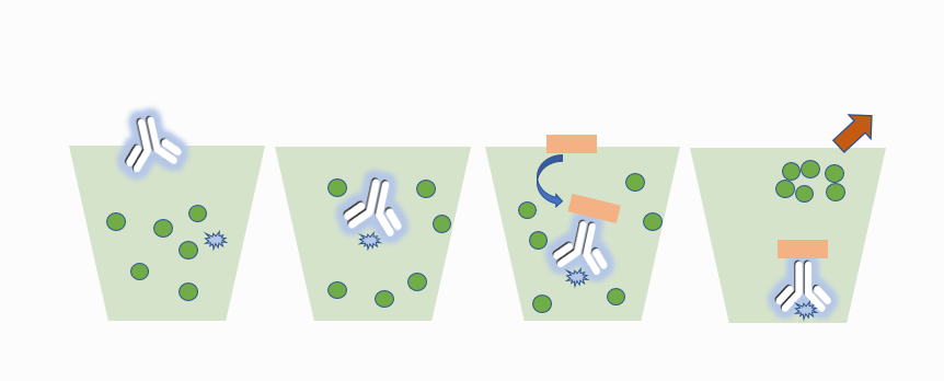
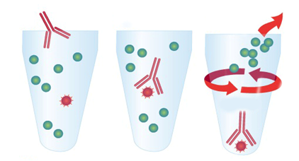
Immunoprecipitation (IP) is a method for purifying and enriching target proteins from specific mixtures (usually cell lysates or expression supernatants) by using antigen-antibody specific reactions. The traditional IP experiment is that after the antibody is combined with the target protein, it is incubated with agarose or magnetic beads coupled with Protein A /G (Fc fragment of binding antibody), and the bead-protein A/G- antibody-target protein complex is obtained by centrifugation. The complex is washed and eluted for subsequent downstream experiments. IP is an important step in many protein-related researches. It is used to study the existence, relative abundance, up-down regulation of protein expression, protein-to-stability and interaction of proteins.


Traditional IP protocol

Summary: The key tools I need to use in the whole
process of IP are,


1) primary antibody: select the primary antibody that can
react with the sample and do IP experiment;


2) Potein APotein G or Protein
A /G
:Potein A, Potein
G and Protein A /G have different binding abilities to immunoglobulins from
different sources and subtypes. how to choose? Please refer to https://www.abbkine.com/affinity-of-protein-a-protein-g-and-protein-ag/


3) Selection Guide:


Cat#

Product name

BMR20500

PurKine™
Protein A Resin

BMR20504

PurKine™
Protein A Resin 4FF

BMR20600

PurKine™
Protein G Resin

BMR20604

PurKine™
Protein G Resin 4FF

BMR20704

PurKine™
Protein A/G Resin 4FF


Step 2: Finding that there is no suitable primary antibody to meet the immunoprecipitation I need to do?


Some target proteins cannot be immunoprecipitated because there is no
corresponding specific antibody. For example, the antigen epitope recognized by
the antibody is shielded by the interaction protein, unable to interact with
the target protein or the antibody selection is inappropriate, the selected
antibody cannot recognize the protein in natural conformation, etc. After an
epitope tag protein is used, the tag antibody against this epitope can be
selected for immunoprecipitation. Manufacturers will introduce some common
label antibodies for IP experiments, involving labels such as flag, HA, His and
Myc to meet the needs of most customers.


In particular, the agarose/magnetic bead coupled labeled antibody can
optimize the immunoprecipitation experiment to the greatest extent and save
time and cost.


1) Principle: The step of Protein A/G binding with antigen-antibody complex is omitted, and the problem of weak binding ability between Protein A/G and antibody is solved. After the conjugated antibody is directly combined with the target protein, the target protein is separated from the cell lysate by centrifugation or using a magnetic rack. The high-specificity monoclonal label antibody can realize high yield and high purity, and the stable and pre-sealed filler and the specific antibody reduce non-specific binding in the immunoprecipitation process.


Agarose/magnetic bead conjugated label antibody is applied to IP

2) Selection Guide:


Cat#

Product name

A02010AGB

Anti-DDDDK Tag
Mouse Monoclonal Antibody (1B10), Agarose

A02010MGB

Anti-DDDDK Tag
Mouse Monoclonal Antibody (1B10), Magnetic Beads

A02040AGB

Anti-HA Tag
Mouse Monoclonal Antibody (4F6), Agarose

A02040MGB

Anti-HA Tag
Mouse Monoclonal Antibody (4F6), Magnetic Beads

A02050AGB

Anti-His Tag
Mouse Monoclonal Antibody (5C3), Agarose

A02050MGB

Anti-His Tag
Mouse Monoclonal Antibody (5C3), Magnetic Beads

A02060AGB

Anti-Myc Tag
Mouse Monoclonal Antibody (2D5), Agarose

A02060MGB

Anti-Myc Tag
Mouse Monoclonal Antibody (2D5), Magnetic Beads


Step 3: How to deal with heavy chain and light chain interference in WB (IP-WB) after immunoprecipitation?


In WB verification after immunoprecipitation experiment, when
conventional Anti-IgG (H+L) enzyme labeled secondary antibody is used, two
bands corresponding to heavy chain (50kDa) and light chain (25kDa) generated
after denaturation of immunoprecipitation primary antibody will usually appear.
If the molecular weight of the detected target protein is near here, it will be
interfered by the two bands. How to avoid interference,


Method 1: Select the secondary antibody that only
reacts with heavy chain or light chain. If the molecular weight of the target
protein is less than 30KD, in order to avoid interference of antibody light
chain, heavy chain specific secondary antibody is selected; If the molecular
weight of the target protein is more than 30KD, in order to avoid interference
of antibody heavy chain, light chain specific secondary antibody is selected;


Selection Guide:


Cat#

Product name

Description

A25012

IPKine™ HRP,
Goat Anti-Mouse IgG LCS

light chain
specific

A25022

IPKine™ HRP,
Mouse Anti-Rabbit IgG LCS

light chain
specific

A25112

IPKine™ HRP, Goat
Anti-Mouse IgG HCS

Heavy chain
specific

A25222

IPKine™ HRP,
Goat Anti-Rabbit IgG HCS

Heavy chain
specific


Method 2: When selecting WB primary antibody, select different species from IP primary antibody to avoid interference from IP primary antibody;


Method 3: Select the WB primary antibody directly coupled with HRP, and omit the
step of secondary antibody.  Immune
precipitation is broken in three steps.


If you have any questions, welcome to contact
us.

Complete solution of immunoprecipitation (IP)-break down the key steps of IP step by step!

Step 1: Understand what immunoprecipitation is? Why should I do
immunoprecipitation?


Immunoprecipitation (IP) is a method for purifying and enriching target proteins from specific mixtures (usually cell lysates or expression supernatants) by using antigen-antibody specific reactions. The traditional IP experiment is that after the antibody is combined with the target protein, it is incubated with agarose or magnetic beads coupled with Protein A /G (Fc fragment of binding antibody), and the bead-protein A/G- antibody-target protein complex is obtained by centrifugation. The complex is washed and eluted for subsequent downstream experiments. IP is an important step in many protein-related researches. It is used to study the existence, relative abundance, up-down regulation of protein expression, protein-to-stability and interaction of proteins.


Traditional IP protocol

Summary: The key tools I need to use in the whole
process of IP are,


1) primary antibody: select the primary antibody that can
react with the sample and do IP experiment;


2) Potein APotein G or Protein
A /G
:Potein A, Potein
G and Protein A /G have different binding abilities to immunoglobulins from
different sources and subtypes. how to choose? Please refer to https://www.abbkine.com/affinity-of-protein-a-protein-g-and-protein-ag/


3) Selection Guide:


Cat#

Product name

BMR20500

PurKine™
Protein A Resin

BMR20504

PurKine™
Protein A Resin 4FF

BMR20600

PurKine™
Protein G Resin

BMR20604

PurKine™
Protein G Resin 4FF

BMR20704

PurKine™
Protein A/G Resin 4FF


Step 2: Finding that there is no suitable primary antibody to meet the immunoprecipitation I need to do?


Some target proteins cannot be immunoprecipitated because there is no
corresponding specific antibody. For example, the antigen epitope recognized by
the antibody is shielded by the interaction protein, unable to interact with
the target protein or the antibody selection is inappropriate, the selected
antibody cannot recognize the protein in natural conformation, etc. After an
epitope tag protein is used, the tag antibody against this epitope can be
selected for immunoprecipitation. Manufacturers will introduce some common
label antibodies for IP experiments, involving labels such as flag, HA, His and
Myc to meet the needs of most customers.


In particular, the agarose/magnetic bead coupled labeled antibody can
optimize the immunoprecipitation experiment to the greatest extent and save
time and cost.


1) Principle: The step of Protein A/G binding with antigen-antibody complex is omitted, and the problem of weak binding ability between Protein A/G and antibody is solved. After the conjugated antibody is directly combined with the target protein, the target protein is separated from the cell lysate by centrifugation or using a magnetic rack. The high-specificity monoclonal label antibody can realize high yield and high purity, and the stable and pre-sealed filler and the specific antibody reduce non-specific binding in the immunoprecipitation process.


Agarose/magnetic bead conjugated label antibody is applied to IP

2) Selection Guide:


Cat#

Product name

A02010AGB

Anti-DDDDK Tag
Mouse Monoclonal Antibody (1B10), Agarose

A02010MGB

Anti-DDDDK Tag
Mouse Monoclonal Antibody (1B10), Magnetic Beads

A02040AGB

Anti-HA Tag
Mouse Monoclonal Antibody (4F6), Agarose

A02040MGB

Anti-HA Tag
Mouse Monoclonal Antibody (4F6), Magnetic Beads

A02050AGB

Anti-His Tag
Mouse Monoclonal Antibody (5C3), Agarose

A02050MGB

Anti-His Tag
Mouse Monoclonal Antibody (5C3), Magnetic Beads

A02060AGB

Anti-Myc Tag
Mouse Monoclonal Antibody (2D5), Agarose

A02060MGB

Anti-Myc Tag
Mouse Monoclonal Antibody (2D5), Magnetic Beads


Step 3: How to deal with heavy chain and light chain interference in WB (IP-WB) after immunoprecipitation?


In WB verification after immunoprecipitation experiment, when
conventional Anti-IgG (H+L) enzyme labeled secondary antibody is used, two
bands corresponding to heavy chain (50kDa) and light chain (25kDa) generated
after denaturation of immunoprecipitation primary antibody will usually appear.
If the molecular weight of the detected target protein is near here, it will be
interfered by the two bands. How to avoid interference,


Method 1: Select the secondary antibody that only
reacts with heavy chain or light chain. If the molecular weight of the target
protein is less than 30KD, in order to avoid interference of antibody light
chain, heavy chain specific secondary antibody is selected; If the molecular
weight of the target protein is more than 30KD, in order to avoid interference
of antibody heavy chain, light chain specific secondary antibody is selected;


Selection Guide:


Cat#

Product name

Description

A25012

IPKine™ HRP,
Goat Anti-Mouse IgG LCS

light chain
specific

A25022

IPKine™ HRP,
Mouse Anti-Rabbit IgG LCS

light chain
specific

A25112

IPKine™ HRP, Goat
Anti-Mouse IgG HCS

Heavy chain
specific

A25222

IPKine™ HRP,
Goat Anti-Rabbit IgG HCS

Heavy chain
specific


Method 2: When selecting WB primary antibody, select different species from IP primary antibody to avoid interference from IP primary antibody;


Method 3: Select the WB primary antibody directly coupled with HRP, and omit the
step of secondary antibody.  Immune
precipitation is broken in three steps.


If you have any questions, welcome to contact
us.

Complete solution of immunoprecipitation (IP)-break down the key steps of IP step by step!

Step 1: Understand what immunoprecipitation is? Why should I do
immunoprecipitation?


Immunoprecipitation (IP) is a method for purifying and enriching target proteins from specific mixtures (usually cell lysates or expression supernatants) by using antigen-antibody specific reactions. The traditional IP experiment is that after the antibody is combined with the target protein, it is incubated with agarose or magnetic beads coupled with Protein A /G (Fc fragment of binding antibody), and the bead-protein A/G- antibody-target protein complex is obtained by centrifugation. The complex is washed and eluted for subsequent downstream experiments. IP is an important step in many protein-related researches. It is used to study the existence, relative abundance, up-down regulation of protein expression, protein-to-stability and interaction of proteins.


Traditional IP protocol

Summary: The key tools I need to use in the whole
process of IP are,


1) primary antibody: select the primary antibody that can
react with the sample and do IP experiment;


2) Potein APotein G or Protein
A /G
:Potein A, Potein
G and Protein A /G have different binding abilities to immunoglobulins from
different sources and subtypes. how to choose? Please refer to https://www.abbkine.com/affinity-of-protein-a-protein-g-and-protein-ag/


3) Selection Guide:


Cat#

Product name

BMR20500

PurKine™
Protein A Resin

BMR20504

PurKine™
Protein A Resin 4FF

BMR20600

PurKine™
Protein G Resin

BMR20604

PurKine™
Protein G Resin 4FF

BMR20704

PurKine™
Protein A/G Resin 4FF


Step 2: Finding that there is no suitable primary antibody to meet the immunoprecipitation I need to do?


Some target proteins cannot be immunoprecipitated because there is no
corresponding specific antibody. For example, the antigen epitope recognized by
the antibody is shielded by the interaction protein, unable to interact with
the target protein or the antibody selection is inappropriate, the selected
antibody cannot recognize the protein in natural conformation, etc. After an
epitope tag protein is used, the tag antibody against this epitope can be
selected for immunoprecipitation. Manufacturers will introduce some common
label antibodies for IP experiments, involving labels such as flag, HA, His and
Myc to meet the needs of most customers.


In particular, the agarose/magnetic bead coupled labeled antibody can
optimize the immunoprecipitation experiment to the greatest extent and save
time and cost.


1) Principle: The step of Protein A/G binding with antigen-antibody complex is omitted, and the problem of weak binding ability between Protein A/G and antibody is solved. After the conjugated antibody is directly combined with the target protein, the target protein is separated from the cell lysate by centrifugation or using a magnetic rack. The high-specificity monoclonal label antibody can realize high yield and high purity, and the stable and pre-sealed filler and the specific antibody reduce non-specific binding in the immunoprecipitation process.


Agarose/magnetic bead conjugated label antibody is applied to IP

2) Selection Guide:


Cat#

Product name

A02010AGB

Anti-DDDDK Tag
Mouse Monoclonal Antibody (1B10), Agarose

A02010MGB

Anti-DDDDK Tag
Mouse Monoclonal Antibody (1B10), Magnetic Beads

A02040AGB

Anti-HA Tag
Mouse Monoclonal Antibody (4F6), Agarose

A02040MGB

Anti-HA Tag
Mouse Monoclonal Antibody (4F6), Magnetic Beads

A02050AGB

Anti-His Tag
Mouse Monoclonal Antibody (5C3), Agarose

A02050MGB

Anti-His Tag
Mouse Monoclonal Antibody (5C3), Magnetic Beads

A02060AGB

Anti-Myc Tag
Mouse Monoclonal Antibody (2D5), Agarose

A02060MGB

Anti-Myc Tag
Mouse Monoclonal Antibody (2D5), Magnetic Beads


Step 3: How to deal with heavy chain and light chain interference in WB (IP-WB) after immunoprecipitation?


In WB verification after immunoprecipitation experiment, when
conventional Anti-IgG (H+L) enzyme labeled secondary antibody is used, two
bands corresponding to heavy chain (50kDa) and light chain (25kDa) generated
after denaturation of immunoprecipitation primary antibody will usually appear.
If the molecular weight of the detected target protein is near here, it will be
interfered by the two bands. How to avoid interference,


Method 1: Select the secondary antibody that only
reacts with heavy chain or light chain. If the molecular weight of the target
protein is less than 30KD, in order to avoid interference of antibody light
chain, heavy chain specific secondary antibody is selected; If the molecular
weight of the target protein is more than 30KD, in order to avoid interference
of antibody heavy chain, light chain specific secondary antibody is selected;


Selection Guide:


Cat#

Product name

Description

A25012

IPKine™ HRP,
Goat Anti-Mouse IgG LCS

light chain
specific

A25022

IPKine™ HRP,
Mouse Anti-Rabbit IgG LCS

light chain
specific

A25112

IPKine™ HRP, Goat
Anti-Mouse IgG HCS

Heavy chain
specific

A25222

IPKine™ HRP,
Goat Anti-Rabbit IgG HCS

Heavy chain
specific


Method 2: When selecting WB primary antibody, select different species from IP primary antibody to avoid interference from IP primary antibody;


Method 3: Select the WB primary antibody directly coupled with HRP, and omit the
step of secondary antibody.  Immune
precipitation is broken in three steps.


If you have any questions, welcome to contact
us.