Review of Related Articles:
Research
Hotspots of Cell Apoptosis: Bcl-2 Monoclonal Antibody
Abbkine
P53 antibody helps basic cancer research!
Marker of myofibroblast antibody-α-SMA/ACTA2 Monoclonal Antibody
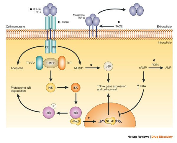
Tumor necrosis factor (TNF, cachexin, or
cachectin; once named as tumor necrosis factor alpha or TNFα) is a cell
signaling protein (cytokine) involved in systemic inflammation and is one of
the cytokines that make up the acute phase reaction. It is produced chiefly by
activated macrophages, although it can be produced by many other cell types
such as CD4+ lymphocytes, NK cells, neutrophils, mast cells, eosinophils, and
neurons. TNF is a member of the TNF superfamily, consisting of various
transmembrane proteins with a homologous TNF domain.
The primary role of TNF is in the regulation of immune cells. TNF, being an endogenous pyrogen, is able to induce fever, apoptotic cell death, cachexia, inflammation and to inhibit tumorigenesis and viral replication and respond to sepsis via IL1- & IL6-producing cells. Dysregulation of TNF production has been implicated in a variety of human diseases including Alzheimer's disease, cancer, major depression, psoriasis and inflammatory bowel disease (IBD). Though controversial, studies of depression and IBD are currently being linked to increased levels of TNF. Recombinant TNF is used as an immunostimulant under the INN tasonermin. TNF can be produced ectopically in the setting of malignancy and parallels parathyroid hormone both in causing secondary hypercalcemia and in the cancers with which excessive production is associated.
Abbkine TNF-α
Polyclonal Antibody helps basic cancer research! Basic information about
the product:
Product name | #CAT | Reactivity | Applications | Dilution ratio | Observed Band(KD) |
TNF-α Polyclonal Antibody | ABP0127 | Human/Mouse/Rat | WB/IF/IHC-P/ELISA | WB (1:500-1:2000) IF (1:200-1:1000) IHC-P (1:100-1:300) ELISA(1/10000) | 16KD |
Abbkine TNF-α Polyclonal Antibody verification results:
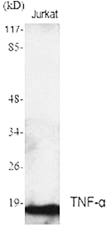 | Western Blot analysis using Abbkine TNF-α Polyclonal Antibody. |
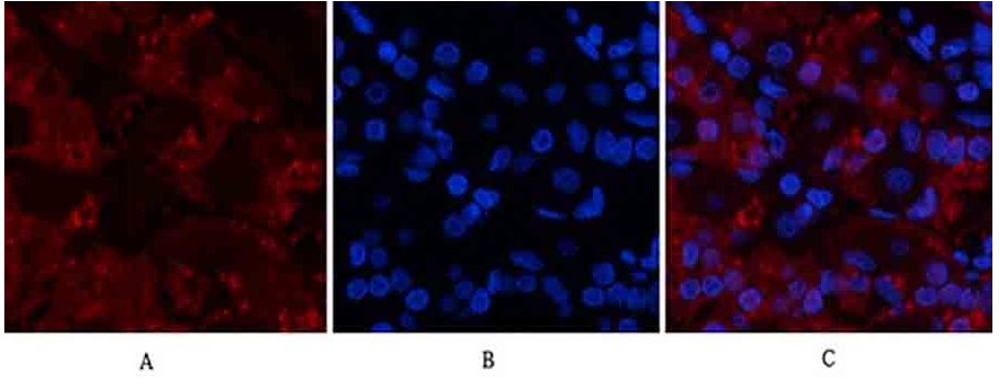 | Immunofluorescence analysis of human stomach tissue using Abbkine TNF-α Polyclonal Antibody (1:200). |
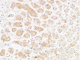 | Immunohistochemical analysis of paraffin-embedded Human stomach (1:100). |